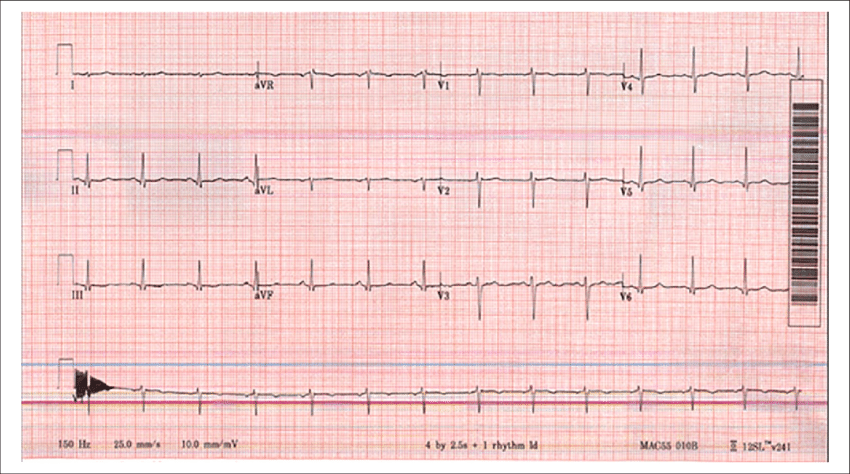
The electrocardiogram (ECG) is a vital diagnostic tool used to evaluate the electrical activity of the heart. One of the key components of the ECG is the P wave, which represents the depolarization of the atria. The P wave is an important parameter to assess the atrial activity and can provide valuable information regarding cardiac function. The P wave is usually analyzed in relation to the QRS complex, which represents ventricular depolarization. The relationship between these two components is known as the P wave axis or PRT axes.

What is Normal PRT Axes?
The normal P wave axis is typically between 0 and +90 degrees. The axis is determined by analyzing the P wave in leads II and III of the ECG. In a normal ECG, the P wave in lead II is usually positive, while the P wave in lead III is usually negative. The P wave axis is the angle at which these two leads intersect, and it can provide valuable information about the location of the atrial depolarization.
When analyzing the P wave axis, it is important to consider the overall context of the ECG. Some factors that can influence the P wave axis include age, gender, and body habitus. In general, the P wave axis tends to shift to the left with increasing age. In women, the P wave axis may be slightly more leftward than in men due to differences in cardiac anatomy. Additionally, factors such as obesity, lung disease, and pericardial effusion can all affect the P wave axis.
Abnormal PRT Axes
Abnormalities in the P wave axis can provide valuable diagnostic information and can help identify underlying cardiac pathology. A leftward shift in the P wave axis (i.e., greater than +90 degrees) is often associated with left atrial enlargement. This can occur in conditions such as mitral stenosis, mitral regurgitation, and left ventricular failure. Conversely, a rightward shift in the P wave axis (i.e., less than 0 degrees) is often associated with right atrial enlargement. This can occur in conditions such as pulmonary hypertension, tricuspid stenosis, and congenital heart disease.
An extreme rightward shift in the P wave axis (i.e., greater than +180 degrees) is known as a “sawtooth” pattern and is often seen in atrial flutter. This arrhythmia is characterized by rapid atrial depolarization, which results in a distinct sawtooth pattern on the ECG. The presence of this pattern can be diagnostic of atrial flutter and can help guide treatment decisions.
Another abnormality of the P wave axis is known as a “pseudo-normal” axis. This occurs when the P wave in lead III appears positive, but the P wave in lead II is biphasic or negative. This can be indicative of underlying pathology such as left atrial enlargement or pulmonary disease.
Conclusion
In summary, the P wave axis is an important component of the ECG that can provide valuable information about cardiac function. The normal P wave axis is typically between 0 and +90 degrees, and abnormalities in the axis can provide diagnostic information about underlying cardiac pathology. When analyzing the P wave axis, it is important to consider the overall context of the ECG, including age, gender, and body habitus. By understanding the significance of the P wave axis, clinicians can more effectively diagnose and manage a variety of cardiac conditions.
